Mobile DR vs. Floor-Mounted Digital X-Ray Systems: UAE Hospital Procurement Guide
Selecting the right digital X-ray machine in the UAE requires an in-depth evaluation of clinical caseloads, diagnostic imaging quality, radiation protection guidelines, physical facility infrastructure, and long-term equipment reliability. In the context of Dubai, Abu Dhabi, and across the Northern Emirates, healthcare leaders face a fundamental architectural choice: whether to deploy mobile digital radiography (DR) units, fixed floor-mounted or ceiling-suspended radiography suites, or a balanced hybrid configuration.
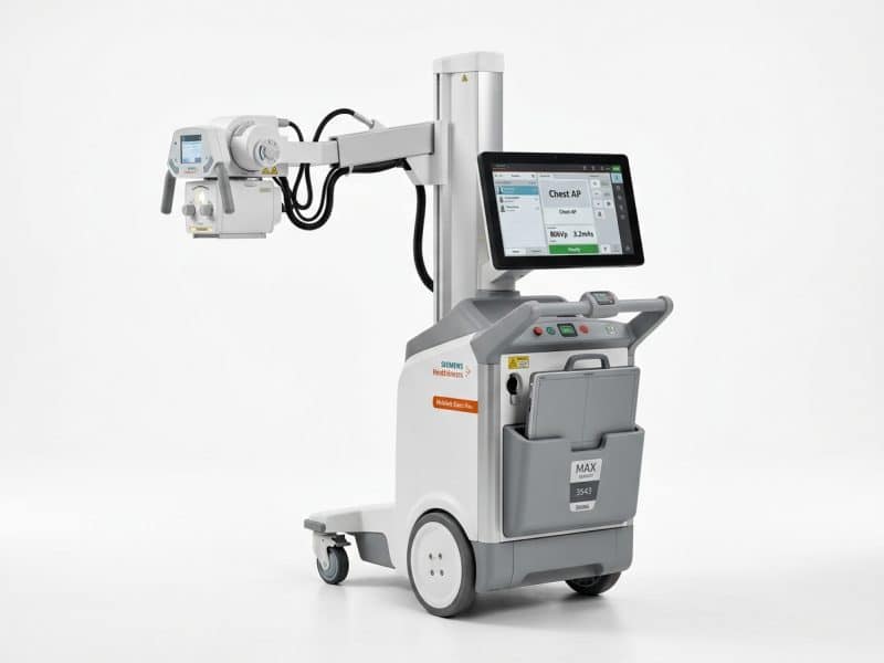
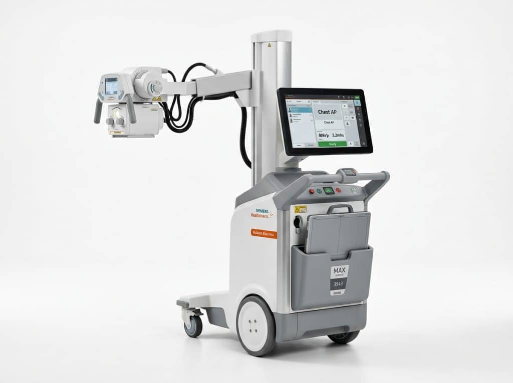
Mobile DR systems deliver immediate point-of-care radiography directly at the bedside in Intensive Care Units (ICU), Neonatal ICUs (NICU), Emergency Departments (ED), operating suites, and isolation wards. Conversely, fixed digital radiography installations provide an optimized, shielded environment designed for high-throughput routine diagnostic exams, complex orthopedic views, and outpatient patient traffic.
Determining the optimal modality mix hinges on patient mobility levels, clinical throughput demands, generator kilowatt output, Flat Panel Detector (FPD) performance, and facility workflow. Ultravision Medical Equipment Trading LLC supports healthcare organizations throughout the UAE with specialized radiology equipment solutions and comprehensive biomedical engineering services.
1. Clinical Comparison: Mobile Bedside vs. Fixed Radiography Suites
Mobile Digital Radiography: Point-of-Care Bedside Imaging
Mobile DR eliminates the clinical risks and logistical delays associated with transporting critically ill or immobilized patients to central radiology rooms. Key clinical applications include:
- Intensive Care Units (ICU): Rapid bedside chest radiography to confirm endotracheal tube placement, central venous catheter positions, and acute pulmonary conditions.
- Neonatal ICUs (NICU): Low-dose neonatal imaging using dedicated micro-exposure pediatric protocols directly inside incubators.
- Emergency & Trauma Resuscitation: Immediate diagnostic radiography for unstable polytrauma patients where transfer to fixed rooms is clinically contraindicated.
- Operating Theatres (OR): Immediate intraoperative imaging during orthopedic pinning, extremity alignment, or surgical sponge/instrument verification.
- Isolation & Infectious Wards: Bedside imaging that prevents cross-contamination across general hospital corridors and outpatient waiting areas.
Motor-driven steering, compact gantry profiles, and wireless flat panel detectors provide agile maneuverability around crowded hospital beds, ventilators, and infusion pumps. However, bedside imaging demands stringent operator scatter control, collimator precision, and strict infection control barriers.
Floor-Mounted and Ceiling-Suspended Digital X-Ray Suites
Fixed digital radiography installations form the backbone of dedicated diagnostic imaging departments:
- Floor-Mounted Systems: The X-ray tube column travels along heavy-duty floor rails or floor-to-wall tracks. They are ideal for facilities with low ceiling heights, existing HVAC duct obstacles, or where structural ceiling slab reinforcement is impractical.
- Ceiling-Suspended Systems: The tube carriage is suspended from an overhead longitudinal and transverse ceiling rail assembly. This configuration maximizes floor space, allows completely unobstructed patient transfer around the four-way elevating radiographic table, and accommodates advanced multi-angle trauma protocols.
- Automated Tracking & Stitching: High-end fixed suites feature motorized auto-tracking between the tube and vertical bucky stand, as well as multi-exposure spine and full-leg image stitching for orthopedic assessments.
Technical Comparison: Mobile DR vs. Fixed Digital Radiography Suites
| Engineering Feature | Mobile Digital Radiography (DR) | Floor-Mounted / Ceiling-Suspended DR |
|---|---|---|
| Installation Footprint | Compact mobile cart with dedicated wall charging bay | Fully shielded radiographic room with separate operator console |
| Generator Power Output | Typically 30 kW – 32 kW (monoblock / capacitor-discharge) | High-frequency 50 kW – 80 kW dedicated generator |
| Examination Throughput | Governed by transit times, elevator transit, and bedside positioning | High volume: 15 to 25+ structured examinations per hour |
| Maneuverability & Reach | Motor-driven mobility across wards, elevators, and corridors | Precision motorized tracking around patient table and wall bucky |
| Flat Panel Detector (FPD) | High-durability wireless portable FPD (typically 35x43 cm) | Dual FPD configuration (fixed bucky + wireless table detector) |
| Primary Clinical Domain | ICU, NICU, emergency resuscitation, OR, and isolation units | Outpatient radiology, spine/pelvis orthopedics, general triage |
| Power Supply Connection | On-board battery bank with standard 13A/15A charging inlet | Dedicated 3-phase 380V/400V electrical power feed |
| Radiation Protection Paradigm | Operator standoff distance (>=2m), mobile shields, TLD monitoring | Engineered room lead lining (mm Pb) and leaded control window |
Hospital Procurement Recommendation: Facilities managing moderate-to-high critical care or surgical patient volumes should deploy mobile DR as a strategic bedside complement—never as an operational substitute for high-power, high-throughput dedicated imaging suites.
2. Flat Panel Detector (FPD) Technology: CsI vs. Gadox
The Flat Panel Detector (FPD) is the primary determinant of radiographic image resolution, low-contrast visibility, and patient radiation dose efficiency. Most indirect conversion digital detectors rely on a scintillator layer coupled to an amorphous silicon (a-Si) thin-film transistor (TFT) array.
Cesium Iodide (CsI) vs. Gadolinium Oxysulfide (Gadox) Scintillators
- Cesium Iodide (CsI): Features a structured columnar, needle-like crystal growth that acts as microscopic fiber-optic light pipes. This structure channels emitted light photons directly to the photodiode layer with minimal lateral light diffusion. CsI delivers superior spatial resolution, lower noise, and dramatically higher Detective Quantum Efficiency (DQE) at lower radiation doses. It is the gold standard for pediatric imaging and high-volume clinical radiography.
- Gadolinium Oxysulfide (Gadox / Gd2O2S): Utilizes a turbid, granular phosphor matrix. While structurally robust and cost-effective, scattered light diffusion within the phosphor layer reduces high-frequency spatial resolution compared to CsI under equivalent exposure levels.
Detective Quantum Efficiency (DQE) and Clinical Dose Optimization
Detective Quantum Efficiency (DQE) quantifies how effectively a detector preserves signal-to-noise ratio from incident X-ray photons to the digital image output. A higher DQE—especially in low-dose exposure ranges (low Kerma)—allows radiology departments to achieve diagnostic clarity while significantly lowering patient radiation exposure, aligning with UAE FANR ALARA requirements. DQE must be evaluated alongside Modulation Transfer Function (MTF), pixel pitch (typically 100 μm to 140 μm), and advanced multi-frequency image post-processing algorithms.
Wireless FPD Engineering for Demanding Mobile Use
Portable detectors used in mobile hospital workflows face demanding mechanical and environmental conditions. Procurement teams should verify:
- Battery Architecture & Hot-Swapping: Independent detector battery modules that allow hot-swapping without shutting down the acquisition software during emergency rounds.
- Ingress Protection & Decontamination: IPX6 or IP54 fluid resistance ratings to tolerate frequent clinical disinfectant wiping without housing degradation.
- Structural Load Bearing: Carbon-fiber casing capable of withstanding substantial uniform surface weight (typically 150 kg to 300 kg) and concentrated point loads for bariatric bedside examinations.
- Drop and Impact Resistance: Military-grade (MIL-STD-810G) drop testing tolerances to withstand accidental bedside falls.
3. UAE Radiation Safety and FANR Compliance
The Federal Authority for Nuclear Regulation (FANR) governs all radiation-producing medical diagnostic equipment in the UAE. Healthcare facilities in Dubai (DHA), Abu Dhabi (DoH), and the Northern Emirates (MOHAP) must secure formal equipment authorizations, maintain radiation protection programs, and establish documented occupational safety protocols before clinical operation.
Fixed X-Ray Room Structural Shielding Assessment
Shielding calculations for fixed radiography rooms must be engineered by a certified medical physicist based on:
- Generator Output & Workload: Maximum tube potential (typically 125 kVp to 150 kVp) and weekly projected exposure volume (mA·min/week).
- Beam Vectors & Use Factors: Primary exposure directions towards the vertical chest bucky and examination table versus scattered radiation trajectories.
- Occupancy Factors of Adjacent Spaces: Unrestricted public waiting areas, administrative offices, corridors, and adjacent clinical rooms.
- Lead Equivalence (mm Pb): Wall lead lining, leaded access doors, and certified lead glass viewing partitions designed to keep dose rates below FANR public and occupational dose constraints.
Mobile X-Ray Radiation Safety Outside Shielded Rooms
Because mobile radiography takes place in unshielded wards and critical care units, strict operational protocols are mandatory:
- Inverse Square Law & Standoff Distance: Operators must utilize exposure cords or wireless triggers enabling a minimum operating distance of 2 meters from the tube and patient.
- Beam Collimation & Scatter Control: Restricting the X-ray beam strictly to the clinical anatomy of interest to minimize lateral scatter.
- Personal Radiation Monitoring: Radiographers and mobile operators must be enrolled in continuous dosimetry programs using Thermoluminescent Dosimeters (TLD) or Instadose badges under FANR oversight.
- Mobile Lead Partitions & Lead Aprons: Providing mobile lead shielding screens for adjacent patients in open ICU bays who cannot be relocated.
4. Site Planning, Power Infrastructure, and Engineering Readiness
Rigorous site readiness planning prevents costly installation delays and ensures long-term operational uptime for both stationary and mobile modalities.
Fixed DR Infrastructure Requirements
- Dedicated Clean Electrical Power: Fixed systems with 50 kW to 80 kW generators require a dedicated three-phase supply (380V/400V ±10%, 50 Hz). Circuit breakers, feeder cables, and protective earthing ( 2 Ohms resistance) must accommodate sudden peak exposure power demands without line voltage drop.
- Overhead Unistrut Ceiling Support: Ceiling-suspended tubes require structurally engineered overhead Unistrut or I-beam grid assemblies rated for static and dynamic gantry loads, perfectly leveled to ensure smooth motorized movement.
- Precision Floor Rail Anchoring: Floor-mounted column rails require level floor slabs (within ±2 mm across the travel span) to eliminate carriage binding and ensure smooth manual or motorized positioning.
- HVAC Thermal Control: Climate control must continuously dissipate heat from high-capacity generators, X-ray tube assemblies, and console computers, maintaining 18°C–24°C and 30%–60% non-condensing relative humidity.
Mobile DR Facility Infrastructure
- Dedicated Charging Bays: Secure parking bays equipped with OEM-compatible 13A or 15A electrical outlets, adequate ventilation for battery charging, and clear access.
- Transit Corridor Clearances: Doorway widths, corridor turning radii, ramp inclines, and elevator dimensions must accommodate the mobile unit's gantry footprint and turning radius.
- Elevator Weight Capacities: Hospital elevators must support the combined weight of the mobile unit (typically 350 kg to 600 kg) plus transport personnel and medical equipment.
- Motor Drive & Anti-Collision Sensors: Powered drive systems with safety bumper interlocks that automatically stop forward motion upon detecting obstacles or pedestrians.
5. PACS Connectivity and Digital Radiography Workflow
Modern digital radiography systems must communicate seamlessly with hospital radiology information platforms to ensure zero demographic errors and rapid clinical reporting.
DICOM Modality Worklist (MWL) & Patient Safety
DICOM MWL enables the acquisition console to pull scheduled patient demographics and procedure identifiers directly from the HIS/RIS. This eliminates manual data entry, prevents misspelled names or swapped medical record numbers (MRN), and ensures accurate study matching in the hospital archive.
DICOM Storage, MPPS & Radiation Dose Structured Reporting (RDSR)
- DICOM Storage & Storage Commitment: Automatically pushes multi-frame and processed radiographic studies to PACS with handshake confirmation before local image deletion.
- Modality Performed Procedure Step (MPPS): Transmits real-time examination status milestones (IN PROGRESS, COMPLETED) back to the RIS.
- Dose Area Product (DAP) & RDSR: Integrates dose-area product meters to record patient radiation exposure and export dose data directly to hospital PACS and national dose registries.
Explore our specialized PACS integration and DICOM workflow services.
Secure High-Speed Wireless & LAN Connectivity
Mobile DR carts rely on enterprise-grade 5 GHz dual-band Wi-Fi (802.11ac/ax) with WPA2/WPA3-Enterprise authentication to transmit high-resolution images instantly from patient bedsides to PACS. Fixed suites utilize redundant 1 Gbps wired LAN drops. Local on-board solid-state storage (SSD) ensures that mobile units continue acquiring exams even if Wi-Fi dead zones are temporarily encountered.
6. Biomedical Maintenance, Quality Assurance, and Calibration
Preventive maintenance is critical to ensure radiation safety, image diagnostic accuracy, and avoid catastrophic component failure. Ultravision Medical Equipment Trading LLC delivers full-lifecycle biomedical maintenance and ISO-aligned calibration services across the UAE.
Critical Preventive Maintenance (PPM) Protocols
- X-Ray Generator Verification: Testing kVp accuracy, mA linearity across varying exposure stations, exposure timer reproducibility, and Automatic Exposure Control (AEC) response.
- Collimator & Optical Field Congruence: Verifying light-field to radiation-field alignment within ±1% of the Source-to-Image Distance (SID) and laser crosshair precision.
- Flat Panel Detector Quality Assurance: Regular dark-field calibration, pixel defect mapping, gain calibration, and phantom image uniformity checks to prevent ring or banding artifacts.
- Mechanical & Electrical Safety: Testing emergency brake interlocks, cable carrier articulation, gantry balancing, grounding resistance, and battery charging health.
Planned Preventive Maintenance (PPM) vs. Corrective Repairs
| Engineering Domain | Planned Preventive Maintenance (PPM) | Corrective Emergency Repairs |
|---|---|---|
| X-Ray Generator | kVp accuracy calibration, timer checks, inverter testing | Inverter board repair, high-voltage tank troubleshooting |
| X-Ray Tube & Housing | Filament emission checks, housing thermal sensor verification | X-ray tube replacement, high-voltage cable replacement |
| Beam Collimator | Light-field to radiation-field congruence calibration | Collimator lamp replacement, shutter motor realignment |
| Flat Panel Detector | Dark/gain calibration, pixel map update, wireless test | Detector interface repair, carbon casing replacement |
| Mobile Drive System | Motor drive testing, electromagnetic brake check, battery test | Drive motor replacement, emergency bumper switch repair |
| Software & Firmware | OEM diagnostic scans, database indexing, safety patch check | Database corruption recovery, OS reinstallation, PACS re-link |
7. Seven-Point Site Readiness and Procurement Checklist
Prior to signing vendor handover certificates and starting clinical patient scanning, hospital biomedical and radiology committees should complete this 7-point verification protocol:
| No. | Verification Milestone | Technical Action Required |
|---|---|---|
| 1 | Clinical Workload Alignment | Confirm whether mobile bedside units, fixed high-throughput suites, or a hybrid model matches clinical case volumes. |
| 2 | Generator & Detector Match | Verify generator kilowatt rating (30–32 kW mobile vs 50–80 kW fixed) and CsI high-DQE detector specifications. |
| 3 | FANR Radiation Safety | Secure FANR equipment import/operation licenses; verify room lead lining (mm Pb) or mobile operator radiation protocols. |
| 4 | Structural Readiness | Validate ceiling Unistrut load capacities, floor rail levelness, elevator weight limits, and corridor pathway clearances. |
| 5 | Electrical & Environmental | Confirm dedicated 3-phase power or 13A/15A charging bays, 2 Ohm clean earthing, and continuous HVAC heat dissipation. |
| 6 | RIS/PACS Connectivity | Complete end-to-end testing for DICOM Modality Worklist, Storage Commitment, RDSR dose logging, and 5 GHz wireless coverage. |
| 7 | Acceptance & PPM Contract | Conduct radiation survey, clinical phantom acceptance testing, radiographer training, and sign comprehensive PPM service contracts. |
8. Conclusion: Engineering-Led Radiography Procurement
Strategic procurement of digital radiography equipment in the UAE demands a balanced engineering approach that harmonizes clinical throughput, radiation safety, civil facility planning, and lifecycle maintenance. While mobile digital radiography provides indispensable point-of-care imaging across critical care wards, fixed floor-mounted and ceiling-suspended suites deliver the high-power reproducibility and high throughput essential for busy outpatient and emergency centers.
Ultravision Medical Equipment Trading LLC partners with healthcare facilities across Dubai, Abu Dhabi, and the UAE, providing vendor-neutral engineering consultancy, turnkey radiology installations, and certified biomedical maintenance services.
To discuss digital radiography procurement, room shielding calculations, or biomedical service agreements, contact our specialized engineering team today.